
Optogenetics-LEDs
Fiber Coupled LEDs for Optogenetics Experiments in Freely Moving Mammals

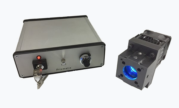
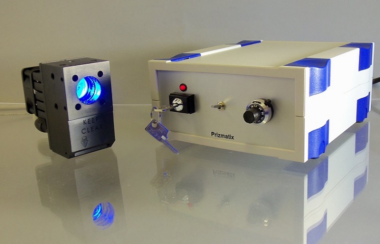
The Prizmatix fiber-coupled Optogenetics-LED modules are specially designed to provide high power and high brightness light to activate or silence various opsins in optogenetics experiments with freely moving mammals.
The typical Optogenetics-LED system comprises Fiber-Coupled Optogenetics-LED, Fiber patch cord, Rotary-Joint (Commutator), thin flexible Optical-fiber (single or dual branch), Sleave and fiber inplant (Cannula).
Optogenetics Toolbox for Freely Moving Mammals Experiments
Prizmatix offers a wide range of standard and customized items comprising the Optogenetics Toolbox. The most useful items for optogenetics experiments in freely moving mammals are:
- Optogenetics-LED
- Extremely low friction Rotary-Joint
- Fiber Optics Cannulae
- Single or Dual Fiber for bilateral activation
- Fiber patch cord
- Pulser for generation of pulse train
For more information on items 2-6 please see Optional Accessories below

Key Features
- High Power density (e.g. at Blue >380 mW/mm²) at implant tip following Rotary-Joint and all fiber connections.
- Unique large and powerful LEDs enable bilateral illumination with single LED and rotary joint for significant cost saving and without compromising power at implant tip.
- Extremely low torque LED-compatible Rotary-Joint suitable for even the smallest animals
- Precisely adjustable power by 10 turns potentiometers (separately for each channel)
- Opto-Isolated TTL Inputs for fast switching (separately for each channel)
- Opto-Isolated Analog Inputs for external power control (separately for each channel)
- Instant warm up time. Rise / Fall time <10µs
Detailed Product Information
| Product P/N | Peak λ [nm] | Cent-roid [nm] | FWHM [nm] | Color Temp [K] | Power LLG3 [mW] | Power LLG5 [mW] | |
|---|---|---|---|---|---|---|---|
| UHP-F-365 | 369 | 372 | 15 | - | 1800 | 4300 | |
| UHP-F-385 | 385 | 387 | 13 | - | 1900 | 3800 | |
| UHP-F-405 | 404 | 406 | 14 | - | 2800 | 6000 | |
| UHP-F-455 | 453 | 456 | 25 | - | 2100 | 4300 | |
| UHP-F-470 | 466 | 468 | 25 | - | 1800 | 5000 | |
| UHP-F-525 | 525 | 528 | 42 | - | 650 | 1300 | |
| UHP-F-545 | 545 | 545 | 98 | - | 2700 | 3700 | |
| UHP-F-560 | 560 | 570 | 97 | - | 1400 | 2100 | |
| UHP-F-625 | 626 | 622 | 20 | - | 1100 | 2200 | |
| UHP-F-630 | 629 | 626 | 22 | - | 1000 | 2000 | |
| UHP-F-730 | 731 | 726 | 35 | - | 750 | 1900 | |
| UHP-F-HCRI | - | - | - | 5700 | 1200 | 2000 | |
| UHP-F-W57 | - | - | - | 5700 | 2300 | 3200 | |
| UHP-F-W65 | - | - | - | 6500 | 2000 | 2800 | |
| UHP-F-WCS | - | - | - | 6500 | 2400 | 3300 | |
| UHP-F-WDS | - | - | - | 5665 | 2500 | 3500 | |
| UHP-F-WSS | - | - | - | 8500 | 2600 | 3700 |
| Accessory Details | Product Photo |
|---|---|
Beam CombinerThe Beam Combiner can join two discrete ICHI LEDs into one output beam, which can then be coupled to a microscope or an optical fiber. For increased versatility, additional optical filters can be installed at the input or the output ports of the Beam Combiner. |
 |
Fiber Coupler AdaptorThe output of ICHI LED or Beam Combiner can be easily changed from collimated output mode to fiber coupled mode. The Fiber Coupler Adaptor (FCA) is suitable for use with high NA optical fibers (optimized for NA 0.5). The FCA enables connection of various fibers and use of ICHI LED system for point in-situ activation. |
 |
Fiber OpticsPrizmatix offers a wide range of standard and customized multi-mode silica / polymer fibers as well as ferrules for optogenetics research. The Polymer Optical fibers are especially useful for neuroscience research as they are extremely flexible and virtually non breakable. The Polymer Fibers typically have high NA well suited for LED light transmission. Prizmatix offers many configurations of fibers including Armored fibers, Y-shaped fiber bundles and much more. |
 |
Rotary JointPrizmatix's Rotary Joint is specially designed for Optogenetics experiments with High NA detachable fibers equipped with FC connectors. The torque required for free movement is very low thus reducing the Rotary Joint's behavioral effect on free moving animals, making it suitable for even the smallest rodents. Prizmatix Rotary Joint can be used with single or multiple output fibers simultaneously, enabling concurrent delivery of light to separate areas of the brain without loss of power or brightness. |
 |
Beam SwitcherThe Beam Switcher allows for simple combining of different light sources to same epi-fluorescent port or to split the light from wide-field illumination to fiber optic in-situ point activation. Typically Beam Switcher shall be used with Fiber Coupler Adaptor (FCA) ad optical fibers. |
|
Filter WheelThe ICHI LED can be equipped with a 6 positions filter wheel at the beam output. This accessory is particularly useful for ICHI-White light source. Prizmatix offers manual and motorized versions of the Filter Wheel. |
|
Microscope AdaptorsThe microscope adaptors enable easy connection of Prizmatix LED light sources to the most widely used microscope brands. Adaptors for epi-fluorescence ports of Nikon, Zeiss, Olympus and Leica microscopes as well as C-mount thread adaptor (1-32 UN 2A thread) are availible. |
|
Liquid Light Guide Adaptor / CollimatorThe output of ICHI LED or Beam Combiner can be easily changed from collimated output mode to light guide coupled mode. The Liquid Light Guide Adaptor (LLG-A) is suitable for use with standard high NA liquid light guides. The Liquid Light Guide Adaptors available for 3mm and for 5mm Light Guides. Same part can be used as fixed collimator on distal end of the liquid light guide. For microscopy applications preferably to use XYZ Collimator (see #11 below) |
|
Liquid Light GuidePrizmatix flexible 3mm or 5mm core liquid light-guide is ideal to conduct light from the large LED emitter to the microscope. The light output from Light Guide equipped with suitable collimator is highly homogeneous and provide flat field illumination. |
|
Liquid Light Guide XYZ CollimatorThe Prizmatix XYZ LLG collimator is an XY and Z adjustable collimator for Liquid Light Guide (LLG) with adaptor for the epi-illumination port of fluorescence microscopes. This collimator can be equipped with Nikon, Olympus, Zeiss or Leica adaptors. |
|
Reference PhotodiodeReference photodiode is useful in order to monitor LED source power over time in experiments required exceptional stability over long time period. The photodiode signal can be used to normalize measurements done over long time period. |
|
Single Dual FiberPrizmatix provides a full solution to the optogenetics in vivo and in vitro fiber optics. Made of silica or polymer fibers, the high NA fibers are assembled to fit any research set-up with various combinations of connectors, ferrules, core diameters and lengths. |
|
Pulser / PulserPlusOptogenetics Pulser / PulserPlus are programmable TTL pulse train generators for pulsing LEDs, lasers and shutters used in Optogenetics activation in neurophysiology and behavioral research. The Pulser device comes with user friendly Windows software that enables easy visual setup of numerous pulse train configurations. |
|
Prizmatix Innovative ApplicationHigh-power LED system allows photostimulation of multiple neurons |
|
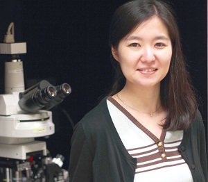
Research led by Dr. Sachiko Tsuda of the Duke-NUS Graduate Medical School in Singapore is directed at better understanding the functional organization of neuron circuits in the brain. She and her colleagues in the lab of Prof. George Augustine recently developed an all-optical approach to study how groups of neurons collectively communicate within brain networks. “Our optogenetic approach enables both the control and detection of the activity of entire populations of neurons simultaneously by using light, which makes it easier to map the connections between different neuron populations,” said Dr. Tsuda, the lead author of their recently published paper. The researchers achieved photostimulation of multiple neurons via the lightsensitive cation channel, channelrhodopsin-2, using the Prizmatix UHP-Mic-LED-460 as the light source and the Andor Mosaic digital micromirror device to create arbitrary spatial patterns of illumination. They simultaneously detected the postsynaptic responses via voltage sensitive dye imaging. “Since voltage-sensitive dye imaging enables observation of synaptic inhibition as well as excitation, we can visualize inhibitory circuits that largely remain to be elucidated,” Dr. Tsuda said. |
|
|
|
|
“The strong output of UHP-Mic-LED-460 solved this problem very effectively. As a bonus, the simple control of its LED via TTL signals also helped us to simplify synchronization of our optical and electrophysiological instruments.” Because they were photostimulating multiple neurons, it was challenging to get enough light power to efficiently activate each neuron. “The strong output of UHP-Mic-LED-460 solved this problem very effectively,” Dr. Tsuda said. “As a bonus, the simple control of its LED via TTL signals also helped us to simplify synchronization of our optical and electrophysiological instruments.” The Prizmatix UHP-Mic-LED-460 provides more than 1.5 W of collimated light, with a peak excitation of 460 nm, and is optimal for photostimulating a large number of neurons. Prizmatix LEDs all feature a direct TTL input for fast switching with a rise/fall time of microseconds, much faster than the millisecond pulses required for optogenetic applications. As a proof-of-principle, the researchers used their technique to control the activity of cerebellar interneurons while simultaneously recording inhibitory responses in multiple Purkinje neurons, which are the postsynaptic targets for the interneurons. The results demonstrated that their all-optical technique allows rapid and quantitative analysis of the spatial organization of neuronal circuits. “We believe that this approach will greatly aid understanding of the functional organization of neuronal circuits,” Dr. Tsuda said. The researchers are now working to increase the resolution of photostimulation mapping by optimizing the detection of neuronal responses, and they plan to try the approach with genetically encoded fluorescent indicators to achieve cell-type specificity for the neuronal responses. Dr. Tsuda adds that their approach could be used to analyze neuronal circuits in intact brains, and for such in vivo applications, the UHP-Mic-LED-460 would be a key component because of its high power and simple control. Research Paper: Tsuda S, Kee MZ, Cunha C, Kim J, Yan P, Loew LM, Augustine GJ.. Probing the function of neuronal populations: combining micromirror-based optogenetic photostimulation with voltage-sensitive dye imaging. Neurosci Res. 2013 Jan;75(1):76-81 Prizmatix Products for Research
In this research Dr. Tsuda used Prizmatix Ultra High Power Collimated LED (P/N: UHP-Mic-LED-460),
the best product for year 2013. Since then we improved UHP product lines and now Prizmatix offers more advanced models.
Currently main product lines are: Advantages of UHP-T series:Location of high current LED driverIn UHP-T-LED the high current LED driver is located inside LED head rather than in controller. In similar products from other vendors the high current driver located in the controller box. When high current flows from controller to LED head high RFI/EMI interference may disturb delicate measurements. In UHP-T-LED models the LED head is grounded and serve as Faraday cage reducing the RFI/EMI interference. Optically Isolated TTL and Analog InputsThe controller of UHP-T-LED features, as standard, TTL input for very fast triggering (or ON/OFF strobing) of LED light without need of a shutter. The Analog input provides simple way to control the LED light power from computer. Both inputs are independent and optically isolated to eliminate ground loops. Low Optical Noise OptionMost modules can be purchased with Low Noise (-LN) option. The low optical noise light source is very important in experiments involving such measurements as cell membrane potential imaging by Voltage-Sensitive Dyes (VSD) (potentiometric dyes). In many preparations the change of Voltage-Sensitive Dye fluorescence signal may be just few percent. If the excitation light source exhibits high intensity fluctuations the small VSD changes may be obscured. UHP-T-LED-LN has intensity fluctuations of less than 0.01% RMS between DC to 1MHz, enabling detection of small changes in Voltage-Sensitive Dye fluorescence intensity. |
|
The contemporary LED light source for VSD imaging is UHP-T-460-EP-LN. This model provides besides powerful illumination low RFI/EMI interference, low optical noise and convenient TTL and Analog inputs. All these features are important in LED light-source for Ephys rig environment. |
|

Looking for a specific or custom solution?